Plantar Fasciitis: Biomechanics, Clinical Stages, and Treatment Pathways

Stabbing heel pain in the morning? We provide specialist Plantar Fasciitis treatment in Singapore using 3D Gait Analysis, Shockwave Therapy, and Custom Orthotics. Book your relief today!

Plantar fasciitis is a degenerative musculoskeletal disorder characterised by micro-tears, collagen breakdown, and chronic thickening of the plantar aponeurosis at its calcaneal insertion. While historically classified as an inflammatory condition (“-itis”), histological evidence confirms it is predominantly an unhealed repetitive strain injury—a fasciosis—characterised by failed tissue repair, hypervascularity, and collagen disorganisation.

The plantar fascia is a dense band of connective tissue running from the medial tubercle of the calcaneus (heel bone) forward to the base of the proximal phalanges. It forms the primary structural tie-rod of the foot, maintaining arch height and facilitating the windlass mechanism. As the big toe extends during propulsion, the fascia tightens around the metatarsal heads, elevating the longitudinal arch and transforming the foot into a rigid lever for forward push-off. When this biomechanical system is chronically overloaded, high tensile strain triggers micro-avulsions at the heel origin, producing the classic focal stabbing pain.

Stop the Heel Pain - Book Appointment
       Resting Plantar Aponeurosis                 Windlass Mechanism Under Strain
   [ Calcaneus ────── Plantar Fascia ────── MTP ]   [ Calcaneus ──▲── High Tensile Strain ──▲── MTP ]
     Relaxed tissue; low tensile load               Toe dorsiflexion tightens fascia; arch lifts.
     during non-weight-bearing rest.                Overload causes micro-tears at calcaneal origin.

Pathological Triggers in Singapore

While anatomical structure sets baseline vulnerability, urban environmental and lifestyle factors in Singapore regularly trigger and perpetuate chronic plantar heel pain:

  • Unforgiving Urban Surfaces: Singapore’s built environment consists almost entirely of hard, shock-resistant concrete, granite footpaths, and polished indoor tile or marble flooring. Walking barefoot at home on ceramic tiles eliminates natural fat-pad cushioning and exposes the calcaneal attachment to harsh ground reaction forces.

  • Biomechanical Overpronation: Flattening of the medial longitudinal arch during mid-stance and propulsion causes the foot to elongate. This elongation places continuous, non-physiological traction on the plantar fascia, pulling at its insertion on the medial heel.

  • Repetitive Deceleration and Running Volumes: High-impact court sports (such as badminton, squash, and tennis) and road running on tarmac impose substantial eccentric loads through the Achilles-calcaneus-fascia complex, exceeding the tissue’s tensile threshold.

  • Inadequate or Flaccid Footwear: Regular use of non-supportive footwear, including thin slip-on sandals, soft canvas shoes, or unsupportive corporate dress shoes with rigid soles, removes arch stability and increases tensile stress on the aponeurosis.

  • Tight Posterior Kinetic Chain: Chronic tightness in the gastrocnemius and soleus (calf muscle complex) restricts ankle dorsiflexion, forcing early heel lift during gait and magnifying tensile forces across the plantar fascia.

Clinical Stages and Urgent Warning Signs

Plantar fasciitis progresses through distinct clinical phases. Identifying the stage is crucial for selecting appropriate interventions and preventing chronic structural deterioration.

Stage Pathological Presentation & Symptoms Clinical Action Required
Stage 1 (Acute / Early Strain) Sharp stabbing pain on the first few morning steps or after sitting; pain warms up and dulls during light walking. Biomechanical offloading, targeted calf stretches, activity modification, supportive footwear.
Stage 2 (Subacute / Established Fasciosis) Persistent aching throughout daily weight-bearing; post-exercise throbbing; morning stiffness lasts >30 minutes. Dynamic 3D gait analysis, customised 3D-printed orthotics, deep soft tissue mobilisation.
Stage 3 (Chronic / Degenerative Breakdown) Constant, debilitating heel pain; structural thickening (>4.5 mm on ultrasound); secondary plantar tear or calcaneal spur formation. Extracorporeal shockwave therapy, structured tissue loading, intensive rehabilitation programme.

About Plantar Fasciitis

When Is It Urgent?

Heel pain typically develops insidiously, but certain red-flag presentations require immediate medical evaluation:

  1. Suspected Acute Fascial Rupture: A sudden, audible “pop” or tearing sensation in the arch accompanied by acute sharp pain, immediate swelling, and widespread plantar bruising (ecchymosis) points to an acute tear of the plantar aponeurosis.

  2. Calcaneal Stress Fracture: Constant, deep, throbbing bone pain that worsens progressively throughout the day and causes severe pain when the sides of the heel are squeezed laterally (positive calcaneal squeeze test).

  3. Systemic Inflammatory Arthropathies: Bilateral, recalcitrant heel pain accompanied by pain in other peripheral joints, morning lower back stiffness, or eye inflammation (uveitis), which may indicate ankylosing spondylitis or psoriatic arthritis.

  4. Neurological Entrapment (Baxter’s Nerve): Radiating pain, burning, tingling, or numbness extending across the plantar heel or into the lateral foot, suggesting entrapment of the first branch of the lateral plantar nerve.

Professional Clinical Assessment: What We Do in Clinic

Addressing chronic plantar fasciitis requires identifying the underlying mechanical drivers rather than just managing symptoms. At Podiatry Quest Singapore, our comprehensive diagnostic protocol includes:

  • Palpation and Localisation: Pinpoint palpation of the medial calcaneal tubercle, central fascial band, and retrocalcaneal regions to distinguish fasciitis from fat pad atrophy, bursitis, or nerve entrapments.

  • Windlass Mechanism Testing: Assessing passive hallux dorsiflexion to determine whether the mechanical windlass response is functional, restricted, or inducing pain.

  • Ankle Joint and Kinetic Chain Mobility: Measuring weight-bearing lunge dorsiflexion to identify gastrocnemius-soleus complex equinus, which directly contributes to fascial strain.

  • 3D Dynamic Gait and Pressure Profiling: Utilising our 3D running gait analysis to evaluate ground reaction force trajectories, excessive pronation rates, and peak midfoot pressure distributions.

How We Help

Treatment Options: Clinical Pathways

Effective podiatric management focuses on reducing abnormal tensile strain while stimulating tissue regeneration within the degenerated fascia.

                           [Diagnostic Assessment]
                                      │
         ┌────────────────────────────┴────────────────────────────┐
         ▼                                                         ▼
[Biomechanical Offloading Pathway]                        [Tissue Regeneration Pathway]
  • Customised 3D-printed orthotics                         • Radial / Focused Shockwave Therapy
  • Kinetic windlass facilitation                           • Controlled heavy-slow resistance loading
  • Plantar fascial strapping                               • Collagen remodelling protocol

1. Extracorporeal Shockwave Therapy (ESWT)

For chronic, recalcitrant plantar fasciitis lasting longer than six to eight weeks, extracorporeal shockwave therapy is an evidence-based, non-invasive intervention.

Shockwave therapy delivers high-energy acoustic pressure waves directly into the chronical insertion of the plantar fascia. This produces targeted microtrauma that stimulates cellular healing:

  1. Neovascularisation: Acoustic waves trigger the expression of vascular endothelial growth factors (VEGF), stimulating new microvascular growth to deliver blood and nutrients to the avascular, degenerate fascia.

  2. Collagen Remodelling: It stimulates fibroblast proliferation, restarting the natural healing cycle to replace disorganised type-III collagen with resilient, linear type-I collagen.

  3. Substance P Depletion: Shockwaves overstimulate and deplete Substance P (a principal pain neurotransmitter), providing immediate analgesia and long-term neuro-modulation.

The treatment protocol typically comprises 3 to 5 weekly sessions. It is performed in-clinic, involves zero anaesthetic or downtime, and allows patients to maintain daily activities throughout the treatment cycle.

2. Customised 3D-Printed Foot Orthotics

Mechanical unloading is essential to give the healing fascia the structural protection it needs during everyday weight-bearing.

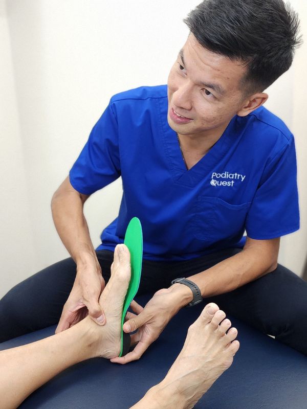
Following 3D laser foot scanning, our podiatrists engineer customised 3D-printed foot orthotics. Unlike generic gel heel cups that merely cushion, medical-grade 3D orthotics mechanically offload the plantar aponeurosis:

  • Contoured Medial Arch Support: Matches the patient’s exact arch height to physically resist arch collapse during mid-stance, reducing peak tensile strain across the central fascial band by up to 30%.

  • First Metatarsal Cut-Outs: Facilitates first-ray plantarflexion, allowing the big toe to extend naturally and engage the windlass mechanism without excessive tension at the heel.

  • Deep Calcaneal Heel Cups: Compresses and centres the natural plantar fat pad directly beneath the calcaneus, restoring the body’s natural shock-absorption system.

Prevention and Daily Foot Health Management

Preventing recurrence requires ongoing management of kinetic chain mobility and foot posture:

  • Avoid Barefoot Walking Indoors: Never walk barefoot on hard tiled or wooden floors at home. Wear contoured, supportive indoor sandals with arch support to prevent repetitive heel trauma.

  • Daily Calf and Fascial Stretching: Perform dedicated gastrocnemius and soleus stretches with your knee straight and bent. Incorporate the Rathleff protocol (high-load, slow resistance calf raises with a rolled towel under the toes) to progressively strengthen fascial load capacity.

  • Opt for Stable, Supportive Shoes: Choose everyday and running shoes with a firm heel counter, torsional rigidity (the shoe should not bend in half), and a moderate heel-to-toe drop (8–10 mm) to reduce strain on the Achilles tendon and plantar fascia.

  • Gradual Training Progression: When returning to running or court sports, adhere to the 10% rule: increase weekly running volume or training intensity by no more than 10% per week to avoid tissue overloading.

Book Your Clinical Consultation at Podiatry Quest Singapore

Stabbing morning heel pain and chronic plantar discomfort should not limit your daily mobility, athletic goals, or quality of life. Our podiatrists provide comprehensive diagnostic evaluations, advanced shockwave therapy, and custom biomechanical solutions to resolve your heel pain definitively.

Contact Podiatry Quest Singapore to schedule your initial heel pain assessment at our Novena, Raffles Quay, Harbourfront, Tampines, or Holland Grove clinics.

🦶 Stop the Heel Pain - Book Appointment

Frequently Asked Questions: Plantar Fasciitis Heel Pain

Can plantar fasciitis cure itself without treatment?

While acute, mild cases may eventually settle with extended rest, chronic plantar fasciitis rarely resolves on its own without correcting the underlying biomechanical overload. Left unaddressed, ongoing micro-tears can lead to chronic fascial degeneration, compensatory gait alterations, and secondary knee, hip, or lower back issues.

Is a heel spur the actual cause of my pain?

No. Heel spurs (calcaneal exostoses) are bony adaptations that form along the tensile pull of the fascia over many years; they are rarely the source of pain. Up to 20% of asymptomatic people have heel spurs, and treating the soft tissue inflammation of the plantar fascia resolves the pain regardless of whether the spur remains.

How many sessions of shockwave therapy will I need?

Most chronic plantar fasciitis cases require a course of 3 to 5 shockwave sessions administered at weekly intervals. Clinical improvements in pain and tissue flexibility are typically felt after the second or third session, with tissue remodelling continuing for up to 12 weeks post-treatment.

Should I continue running or playing sports while recovering?

During the acute, irritable stage, high-impact running, jumping, and court sports should be temporarily replaced with low-impact cardiovascular cross-training, such as swimming, cycling, or rowing. Once morning pain subsides and tissue capacity is rebuilt through custom orthotics and progressive loading, a structured return-to-run programme can begin.

Visit Us

All are welcome!

Podiatry Quest – Holland Grove

Appointment basis; please reach out!

We are situated in a mixed-used estate called Parksuites. There is on-site parking if required.

Bus 92 from Buona Vista MRT.

Monday: Closed
Tuesday: 9am – 8pm
Wednesday: 9am – 6pm
Thursday: Closed
Friday: 9am – 6pm
Saturday: 1230pm – 430pm
Sunday & Public Holidays: Closed

Podiatry Quest – Novena

Appointment basis; please reach out!

The clinic is located on level 3 Novena Square, down a corridor right opposite the lifts.

Monday: Closed
Tuesday: Closed
Wednesday: 9am – 6pm
Thursday: 10am – 7pm
Friday: Closed
Saturday: 9am – 1pm
Sunday & Public Holidays: Closed

Podiatry Quest – Raffles Place

Appointment basis; please reach out!

We are next to Hong Leong building. There is on-site parking in the building if required.

Monday: 9am – 6pm
Tuesday: Closed
Wednesday: 9am – 6pm
Thursday: Closed
Friday: Closed
Saturday: Closed
Sunday & Public Holidays: Closed

Podiatry Quest – Harbourfront

Appointment basis; please reach out!

You’ll be required to register at the lobby with the building concierge and they will direct you to the correct lifts to our clinic – please cater time for this.

Monday: Closed
Tuesday: 9am – 6pm
Wednesday: Closed
Thursday: 9am – 6pm
Friday: Closed
Saturday: 9am – 1pm
Sunday & Public Holidays: Closed

Podiatry Quest – Tampines

Appointment basis; please reach out!

Monday: 9am – 6pm
Tuesday: Closed
Wednesday: Closed
Thursday: Closed
Friday: 9am – 6pm
Saturday: Closed
Sunday & Public Holidays: Closed

Contact Us

Have any questions or inquiries? Get in touch with us today here or on WhatsApp!

Name